Macular Degeneration Foundation
EyeSight.org

Brolucizumab and Retinal Vasculitis

The intraocular inflammation that may occur after intravitreal therapy with brolucizumab (Beovu) can also be accompanied by retinal vasculitis severe enough to cause profound loss of vision, researchers have found.
Source: American Academy of Ophthalmology (AAO). Linda Roach: EyeNet Magazine July 2020
Real-world outcomes. This retrospective analysis of retinal vasculitis in 15 eyes of 12 patients from 10 U.S. centers was the first case series published in a peer-reviewed journal since isolated reports of brolucizumab-associated problems began emerging earlier this year.
The patients’ mean visual acuity (VA) before treatment with brolucizumab was 20/53. By the time retinal vasculitis was diagnosed, it was 20/191 (range, 20/25 to 20/1,600). And at a mean of 25 days following diagnosis and treatment, it was 20/136. Nine eyes (60%) lost 3 lines or more, and five eyes (33%) had VA of less than 20/200.
The vasculitis and intraocular inflammation noted in these eyes ranged from “peripheral vasculitis to occlusion of large retinal arteries around the optic nerve or macula with severe vision loss,” the researchers said. All 12 affected patients were women, which suggests that autoimmunity may be a factor, said coauthor Scott D. Walter MD, MSc, at Retina Consultants in Hartford, Connecticut.
Insidious onset. These adverse outcomes occurred in a pattern distinct from anything ever seen with other approved anti-VEGF drugs, said Dr. Walter, also at the University of Connecticut School of Medicine in Farmington. Specifically, the inflammation associated with brolucizumab “tends to be milder in its early stages and more insidious in onset,” he said. “The patient might not become symptomatic for several weeks after the injection, and the inflammation may be mild enough that patient wouldn’t think to call the office.”
In some patients, “the inflammation was picked up when they returned for a scheduled injection,” Dr. Walter noted. In others, he said, “It was overlooked because there was no intense vitritis or hypopyon.” These are typical signs of intraocular inflammation associated with other anti-VEGF drugs, with onset typically in the first week after injection, he said.
Additionally, “There were multiple exposures to the drug in some cases, and a delay of weeks, as opposed to days, before the onset of clinically apparent intraocular inflammation and retinal vasculitis,” Dr. Walter said. “And if you miss catching this, then it can really get you into trouble.”
If you use brolucizumab. Retina specialists should be alert for inflammation and other events when using brolucizumab, the study authors said. And while researchers try to discover the mechanism behind the problems, Dr. Walter said that he has decided against starting his patients with age-related macular degeneration on brolucizumab, and that he is encouraging those already on it to switch to another anti-VEGF agent.
But for those clinicians who do use the drug, Dr. Walter advises a complete examination of both the anterior and posterior segments to evaluate for subtle signs of inflammation—even for apparently asymptomatic patients—before each subsequent injection. “The most important thing for anyone treating these patients is to not reinject an eye that has active inflammation with brolucizumab or any other anti-VEGF drug.”
How to Wear a Surgical Face Mask

We are all wearing face masks of one kind or another these days. If it’s not bad enough to have limited central vision, some of us also wear glasses that tend to fog up when using a medical procedure mask. Fortunately, Dr. Yang has published a video that may solve that problem for us. It’s intended for medical students, but I’m sure we can pick up a few practical pointers.
“Hands Off” Environmental Controls
by Keith Colgan – Macular Degeneration Foundation
Call it “research” for the Foundation … or just plain laziness, but for the past two years my wife and I have been using a device that allows us to control much of our environment without lifting a finger. Good vision is not a problem for me, but good balance is. So when I enter a room at night, the ability to turn on a light with a simple voice command is important for my safety.

Multiply my needs by a thousand for those who are blind or live with the challenges of low vision. Here are two video’s that provide a good idea of what the Echo Show and Echo Dot by Amazon can do for you.
The AMSLER GRID

Written by: Liz Trauernicht – MDF President
Edited by: Joe L Fontenot MD
What is an AMSLER GRID?

The Amsler Grid is basically a square of horizontal and vertical lines. According to one source, a similar design may have been used in the late 1800’s. But the Amsler Grid we know today gets its name from Marc Amsler, a Swiss ophthalmologist who began promoting its use in 1947.
When used properly, it can help the user to detect visual distortions and loss of vision caused by such diseases as macular edema, central serous chorioretinopathy (CSC) and age-related macular degeneration (AMD).
Early detection of macular disorders is very important … especially today when effective treatments are available when caught early in the process. The Amsler Grid has helped people with macular disease to identify changes in their condition and report it to their eye doctor for evaluation.
What Forms Do Amsler Grids Take?
1. Amsler Grids on paper are the most common form and are available from your doctor and many sources over the Internet. In an attempt to improve accuracy, some use colored backgrounds and lines, while others use a special number of lines and line spacing.
2. Amsler Grid “Apps” are also available for mobile devices like your tablet or cell phone. These are just three that can be downloaded:
For All Devices:
Blindspot Amsler Grid ™ – Download and save for use with any cell phone or tablet. (instructions)
Android Specific:
https://play.google.com/store/apps/details?id=com.ossibussoftware.amsler
Apple Specific:
https://apps.apple.com/hu/app/eyecare-amsler-grid-eye-test/id1016180237
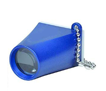
3. Portable viewer: We evaluated a portable key chain “viewer”. It is a novel approach, but we found it to be imprecise and subject to misinterpretation.
4. If you prefer using your computer monitor to check your vision, we have a specialized Amsler Grid called the “Blind Spot Amsler™” available using this link.
How to Use the Amsler
(American Academy of Ophthalmology)
Limitations of the AMSLER GRID
Because the Amsler Grid has limitations, some eye care professionals do not recommended it as a way to “self-assess” disease progression. Here are some of the reasons why:
1. It requires reasonably good near vision to discern the grid lines.
2. Poor patient compliance. Meaning, not everyone is willing or able to maintain a good routine of self-assessment.
3. It is a “subjective” test and prone to misinterpretation.
4. When compared to the tools available to eye doctors, the Amser Grid detects only about one-half of the areas in the visual field where vision is absent or deficient.
5. The eyes and brain can play tricks on you. One of these is called “Perceptual Completion”. This is when the brain fills in or completes gaps in the visual field of each eye. Another challenge is called “Central Fixation”, which causes visual confusion when looking at multiple lines. Perceptual completion and central fixation can make the Amsler Grid unreliable.
Specialized Amsler Grids
A search of the Internet will find references to these less common, high-tech Amsler Grids.
* Threshold Amsler Grid (TAG)
* Accelerated Amsler Grid
* Deformable Amsler Grid
* Three-dimensional Computer-automated Threshold Amsler Grid (3D-CTAG)
These (and other) modified Amsler Grids are attempts to improve the test for use at home between routine eye examinations. Progress is being made in the area of computer and tablet based software to identify macular disorders. All, however, have limitations that make regular visits to your optometrist, ophthalmologist and/or retinal specialist the best way to monitor your vision.
Summary
1. First and foremost, have your eyes examined on a regular basis. How often depends on your age, risk factors and whether you currently wear eyeglasses or contact lenses. Most eye care professionals recommend a comprehensive test every year or two.
2. If you notice a change in your vision, contact an eye care professional immediately.
3. If you have a diagnosis of macular degeneration, an AMSLER GRID may be useful as a “follow-up” tool for monitoring changes in your central vision between eye exams. However, it should never be used to screen yourself for macular disease. Leave that to a trained professional.
“Pitching” Blue Light Protective Eyewear

The Macular Degeneration Foundation published a Special Edition of its Newsletter in July of 2019. It detailed ten common misconceptions regarding macular degeneration. One was the idea that cell phones, televisions and computer screens damage the eyes with “blue light”. The truth is “no scientific evidence has yet revealed that light from such devices causes eye damage”.
The following video, produced in November of 2019 by the Canadian Broadcasting Corporation, interviews Dr. Sunir Garg, an eye surgeon and spokesman for the American Academy of Ophthalmology.
Macular Degeneration Foundation Supports VIS Group

Crossville Chronicle – October 2019: Crossville. Tennessee residents first met Liz Trauernicht, President of the Macular Degeneration Foundation (MDF), when she spoke at the 2014 VIS (Visually Impaired Support Group of Cumberland County) Group’s biennial InSight on Vision. She gave a rousing demonstration of her care for those afflicted with vision loss, especially those diagnosed with AMD.
While Liz was staying in Crossville, she visited the Fair Park Senior Center, bringing info about AMD and leading a lively discussion. Linda Simmons shares, “Since 2014, VIS has been most fortunate to have received tremendous support and loyalty from Liz and the MDF. We can’t be more earnest relating to our community how the MDF has been so important to the VIS Group’s ability of providing important programs for the blind and visually impaired; through the VIS On-Loan Equipment program, the printing of our Resource Guide, monthly support meetings, InSight on Vision, community programs, and monetary support of the TN Council of the Blind and the TN Organization of the Deaf/Blind.”
How smoking can harm your eyes

Statistics have shown that only 1 out of 5 smokers actually are aware of the risk smoking has on their eye health. The following information may convince folks that breaking the habit is imperative to insure a long and healthy life!
1. Cigarette smoke contains toxic chemicals that can irritate and harm the eyes. For example, heavy metals, such as lead and copper, can collect in the lens – the transparent bit that sits behind the pupil and brings rays of light into focus – and lead to cataracts, where the lens becomes cloudy.
2. Smoking can make diabetes-related sight problems worse by damaging blood vessels at the back of the eye (the retina).
3. Smokers are around three times more likely to get age-related macular degeneration – a condition affecting a person’s central vision, meaning that they lose their ability to see fine details.
4. Smokers are 16 times more likely than non-smokers to develop sudden loss of vision caused by optic neuropathy, where the blood supply to the eye becomes blocked.
Criteria for Cataract Surgery

Philip C Hessburg MD
What are the indications for cataract surgery? In short: One should consider having cataracts removed when one’s vision has fallen to levels where he/she cannot do the things one needs – or wants – to do.
If a Delta pilot’s vision drops a line or two, perhaps to 20/25 on the chart due to advancing cataract, it is time for surgery if he /she wants to maintain flying status. While my very elderly illiterate European-born grandmother, whose main joy in life was knitting mittens for 37 Minnesota-based grandchildren, didn’t need cataract surgery though she was probably legally blind (i.e. 20/200 or lower), she was happy knitting mittens.
But what if the patient also has AMD? For many years, ophthalmologists delayed surgery almost until the patient was blind before surgery. Today that is not the case. Today the indications for surgery are no different for patients with or without macular degeneration.
Why the difference?
The difference lies in the revolution which has occurred in cataract surgery over the past 50 years. In the early years of cataract surgery with intraocular lens implantation, the surgery itself was so traumatic that it frequently was associated with profound post-operative inflammation which would – and did – worsen the macular degeneration by inducing inflammatory changes throughout the eye.

Modern cataract surgery, as practiced today by our many fine American cataract surgeons, is so atraumatic that the eye barely knows it has been operated on. In days gone by the incision in the eye was 12-15 mm in length and required six or ten sutures to adequately close. Today cataract surgeons work through a 1.5 mm incision, usually requiring NO sutures to affect a watertight closure.
So today even patients with far advanced macular degeneration are advised to have their cataracts removed. And, although there may be no measurable improvement in the measured central vision on the wall chart, these patients are often among the happiest and most grateful because of their improvement in color vision, in peripheral vision, and in overall “performance vision”. Such patients are also far less prone to stumbles and falls after such surgery.
In summary, forget the old guidelines. Today, cataract surgery often makes a tremendous difference even in patients with far-advanced macular degeneration.
Modern cataract surgery truly can be a modern miracle.
So what does it mean to be blind?

by Philip C Hessburg MD
Well, literally, total loss of all light perception is absolute blindness. That patient would have no perception of light, would not know whether the sun has come up. Fortunately, perhaps as few as 10 or 15% of all people who qualify as “blind” are absolutely blind The rest have levels of sight which range from the ability to perceive light up to levels which would qualify as “legally blind”.
Legal blindness, that level which would qualify the patient for social and government purposes and benefits as “blind”, is usually determined to be 20/200 or below. With that level of vision a person may not be able to read, or drive, or recognize faces very well, but can still get around the house and in society at a level which many folks would not immediately recognize as “that person is blind”. Most patients at that level of vision, that is who are legally blind, do not usually need or use a white cane or a leader dog. This includes, at least in my experience, most patients with far advanced macular degeneration.
Thus, it is important for people with macular degeneration to know that this disease does not result in absolute blindness. At its most profound state, and with modern medical management today this is increasingly rare, age related macular degeneration knocks out the central vision, (that from the macula or center of the retina), and the patient retains peripheral or “side vision”.
If one thinks of this in terms of letters on the eye chart on the wall, macular degeneration may knock vision down to 20/300 or even lower but never cause total loss of vision. (20/300 would mean that this patient would have to walk up to within 20 feet of the chart on the wall to read what the normal person could read from three hundred feet.)
So, in summary , though age related macular degeneration remains a very serious problem, it is increasingly manageable by our gifted ophthalmologists who specialize in retinal diseases. And, unless the patient is also afflicted with some other concurrent serious eye problem (such as glaucoma) the patient will not go totally blind.
New Study Reveals Benefits of Augmented Reality Glasses

Source: Mark Humayun, MD, PhD, director of the USC Dr. Allen and Charlotte Ginsburg Institute for Biomedical Therapeutics, codirector of the USC Roski Eye Institute and University Professor of Ophthalmology at the Keck School of Medicine.
In a new study of patients with retinitis pigmentosa, an inherited degenerative eye disease that results in poor vision, researchers have found that adapted Augmented Reality (AR) glasses can improve patients’ mobility by 50% and grasp performance by 70%.
“Patients with retinitis pigmentosa have decreased peripheral vision and trouble seeing in low light, which makes it difficult to identify obstacles and grasp objects. They often require mobility aids to navigate, especially in dark environments,” said Anastasios N. Angelopoulos, study project lead in Humayun’s research laboratory.
“Currently, wearable low vision technologies using virtual reality are limited and can be difficult to use or require patients to undergo extensive training. Using a different approach — employing assistive technology to enhance, not replace, natural senses — our team adapted AR glasses that project bright colors onto patients’ retinas, corresponding to nearby obstacles,” Humayun said.
Patients with retinitis pigmentosa wore adapted AR glasses as they navigated through an obstacle course based on a U.S. Food and Drug Administration–validated functional test. Using video of each test, researchers recorded the number of times patients collided with obstacles, as well as the time taken to complete the course. Patients averaged 50% fewer collisions with the adapted AR glasses.
Patients also were asked to grasp a wooden peg against a black background — located behind four other wooden pegs — without touching the front items. Patients demonstrated a 70% increase in grasp performance with the AR glasses.
How the AR system works

The AR system overlays objects within a 6-foot wireframe with four bright, distinct colors. In doing so, the glasses provide visual color cues that help people with constricted peripheral vision interpret complex environments, such as avoiding obstacles in dimly lit environments.
To accomplish this, researchers used a process called simultaneous location and mapping, allowing the AR glasses to fully render the 3D structure of a room in real time. The glasses then translated this information into a semitransparent colored visual overlay, which highlighted potential obstacles with bright colors to help patients with spatial understanding and depth perception.
“Through the use of AR, we aim to improve the quality of life for low vision patients by increasing their confidence in performing basic tasks, ultimately allowing them to live more independent lives,” Angelopoulos says.
According to Humayun, while major cost and technical issues remain, this type of assistive technology could eventually become more practical for everyday use in the near future.
=================
Common Misconceptions Regarding Macular Degeneration

by Dan Roberts
MD Foundation Resource Director
888-866-6148
Age-related macular degeneration (AMD) is a progressive disease of the retina wherein the light-sensing cells in the central area of vision (the macula) stop working and eventually die. The disease is thought to be caused by a combination of genetic and environmental factors, and it is most common in people who are age sixty and over.
AMD can be a confusing diagnosis. With so many facets to its diagnosis, symptoms, pathology, and treatment … misunderstandings flourish. Here are ten of the most common misconceptions, each followed by a straight explanation based upon current knowledge.
Misconception #1: “AMD causes blindness.”
Truth: At its worst, AMD will damage only the center of the retina at the back of the eye. This area, the macula, comprises less than 5% of the total retina, but it is responsible for about 35% of the visual field. That means that a person with both eyes affected will eventually find it difficult or impossible to read, drive, or recognize faces.
The peripheral vision, however, is left untouched, so macular degeneration does not, by itself, lead to blindness. Many affected people move about with little or no assistance and lead independent, productive lives. The most successful of them have also learned to use a wide variety of assistive devices such as magnifiers, special bioptic glasses, navigation software, and electronic readers to maximize their peripheral vision and other senses.
Link: What is Macular Degeneration?
Misconception #2: “AMD is a growing epidemic.”
Truth: Recent research has found that the risk of developing AMD has been dramatically lessening over three generations. For that matter, Baby Boomers (born between 1946 and 1964) may experience better retinal health over a longer period of time than the two previous generations. The Baby Boomers and the new Generation X populations are already seeing comparable declines in AMD incidence, attributed possibly to better environmental conditions, sanitation, nutrition, and prevention strategies.
That said, the number of people with visual impairment or blindness in the United States is still expected to double by the year 2050. This is due, though, to the aging population increase, not by any dramatic escalation of the disease risks.
Misconception #3: “Wet and dry AMD are separate diseases.”
Truth: Dry AMD is distinguished by yellowish deposits of cellular debris (“drusen”) in the retina. The material comprising drusen is usually carried away by the blood vessels, but that ability is diminished in AMD.
About 10-15% of dry AMD cases progress to the “wet” form, in which immature blood vessels grow and leak into the retinas of people who have a high genetic inflammatory response. Inflammation is the body’s way of trying to deliver nutrition to injured or diseased tissue. The process is beneficial to the rest of the body, but it can cause scarring and central vision loss if not treated in time.
Therefore, Wet AMD is normally an adverse result of dry AMD, not a separate disease state. People with a normal inflammation response are usually not affected.

Misconception #4: “Reading in dim light will make AMD worse.”
Truth: “Turn on the light”, said Grandma. “You’re going to ruin your eyes.” She meant well, but her suggestion should have been simply, “Turn on the light. You’ll be able to see better.”
Eyes are damaged no more by reading in dim light than are ears by listening to soft music. Actually, the wearing demand on the sight cells increases as the light grows brighter, which may prove harmful to the vision of people with retinal deficiencies. The wisest approach would be to compromise between “enough light to see by” and “too much light.”

Misconception #5: “Viewing cell phone, television, and computer screens damages the eyes.”
Truth: No scientific evidence has yet revealed that light from such devices causes eye damage. The sun and full spectrum lamps which imitate the sun’s high blue content are the two strongest and most harmful sources of light.
By comparison, blue light intensity from cell phones, television, and computer screens is much less than either of those sources. However, until we have more evidence, it may be prudent to simply follow sensible practices like limiting screen time, taking periodic breaks, and taking advantage of light-filtering options.
Link: Blue light research study results
Misconception #6: “Cataract surgery causes AMD”
Truth: The retina is located in the interior of the back of the eye, and cataract surgery replaces the lens at the front of the eye. For that reason, most retinal surgeons say that there is minimal danger of retinal complications from such surgery.
Cataract surgery will not restore vision lost from retinal disease, but replacement of a clouded lens can significantly improve remaining vision, while offering the doctor a clearer view of the retina. In light of the small risk, standard practice is to defer cataract surgery until vision loss from a cataract significantly reduces a patient’s quality of life. At that point, the benefit/risk ratio is sufficiently high to warrant the procedure.
Link: More information concerning Cataract Surgery & AMD
Misconception #7: “Stem cell replacement can cure AMD”
Truth: The media has been full of news about stem cell therapy as a future treatment for AMD. It is true that, in trials, stem cells are replacing the retinal pigment epithelium (RPE) layer that supports the sight cells (photoreceptors). And it is true that scientists are now beginning to replace damaged photoreceptor cells in animal models.
As exciting as this is, stem cell replacement will not be a cure for AMD. Like a patch on a tire, it can restore vision for a time (maybe even until the end of life) but it does not address the underlying cause of the disease. The cure will more likely come from the field of gene replacement therapy, which is still several years down the road.
Link: More information on Stem Cell Therapy
Misconception #8: “Anti-VEGF drugs for wet AMD will reverse vision loss.”
Truth: The anti-VEGF (antiangiogenic) drugs for treatment of wet AMD are designed only to block new blood vessel growth. The intent is not to restore vision, but to maintain current vision and prevent future damage. Some patients, however, do see an improvement after initial injections, but that is mostly due to diminished swelling of the retina and gradual dissipation of collected blood.
While anti-VEGF drugs can effectively stop rapid vision loss from uncontrolled blood vessels, most patients with wet AMD will continue to experience a gradual decline in vision over months and years until new treatments for geographic atrophy (advanced dry AMD) are available. Such treatments are now in trials.

Misconception #9: “Special glasses, eye exercises, electrical stimulation, acupuncture, and nutritional supplements can reverse AMD.”
Truth: Nothing has yet been developed that will reverse AMD. Special prismatic lenses can redirect the wearer’s focus onto a healthier part of the retina. Magnification can enlarge an image to where it can be seen better peripherally. Eye exercises, electrical stimulation, and acupuncture can improve blood flow, temporarily improving visual acuity. And certain nutritional supplements can help to slow the progression of the disease. But once the retinal cells have begun to show the effects of aging, no lens, device, or vitamin can reverse AMD.

Misconception #10: “Nothing can be done”
Truth: By saying that nothing can be done about AMD, a doctor is saying that there is nothing medically that can be done other than anti-VEGF treatment for the wet form. AMD is incurable at this time, but hard-working researchers are close to providing answers. Meanwhile, there is much than can be done to maintain a person’s quality of life with visual impairment. Low vision rehabilitation can provide a strong foundation of knowledge and skills. Assistive devices and computer software equip low vision people with nearly every possible substitute for lost vision. And patient support organizations are ready to provide information and helpful social contact with others who share similar experiences.
Tablet System for those with Low Vision

The MagniLink TAB is a new tablet-based system by LVI Low Vision International. It is “pricey” at over $4000, but claims to offer high performance and user-friendly operation.
The Most Beautiful Flower

by Cheryl L. Costello-Forshey
The park bench was deserted as I sat down to read
Beneath the long, straggly branches of an old willow tree.
Disillusioned by life with good reason to frown,
For the world was intent on dragging me down.
And if that weren’t enough to ruin my day,
A young boy out of breath approached me, all tired from play.
He stood right before me with his head tilted down
And said with great excitement, “Look what I found!”
In his hand was a flower, and what a pitiful sight,
With its petals all worn — not enough rain, or too little light.
Wanting him to take his dead flower and go off to play, I faked a small smile and then shifted away.
But instead of retreating he sat next to my side
And placed the flower to his nose and declared with overacted surprise,
“It sure smells pretty and it’s beautiful, too.
That’s why I picked it; here, it’s for you.”
The weed before me was dying or dead.
Not vibrant of colors, orange, yellow or red.
But I knew I must take it, or he might never leave.
So I reached for the flower, and replied, “Just what I need.”
But instead of him placing the flower in my hand,
He held it midair without reason or plan.
It was then that I noticed for the very first time
That weed-toting boy could not see: he was blind.
I heard my voice quiver, tears shone like the sun
As I thanked him for picking the very best one.
“You’re welcome,” he smiled, and then ran off to play,
Unaware of the impact he’d had on my day.
I sat there and wondered how he managed to see
A self-pitying woman beneath an old willow tree.
How did he know of my self-indulged plight?
Perhaps from his heart, he’d been blessed with true sight.
Through the eyes of a blind child, at last I could see
The problem was not with the world; the problem was me.
And for all of those times I myself had been blind,
I vowed to see the beauty in life, and appreciate every second that’s mine.
And then I held that wilted flower up to my nose
And breathed in the fragrance of a beautiful rose
And smiled as I watched that young boy, another weed in his hand,
About to change the life of an unsuspecting old man.
Speak! Text Reader App for Android

Text readers for the vision impaired are now commonly available for desktop computers. As the technology improves, new mobile apps for cell phones are becoming more powerful and therefore more useful.
Speak! is a brand new one (as of this date) that’s available for Android devices on the Google Play Store. Currently in “Beta”, which means it’s still under development, Speak! performed beautifully on my Samsung Model S8.
Here are a few of its features:

Automatically detects relevant text in an image
Automatically detects the language
Supports very small and far away text
Captured text may also be viewed and enlarged with various background colors
If you give it a try, please let us know how you used it and whether it met your expectations.
“Which is better … 1 or 2?”

If you have been to an eye care provider because you wanted to know if glasses would help you to see better, you’ve probably been asked this question … more than once.
This question is asked while performing a refraction and it can be a nerve racking question. You feel like you don’t want to give the wrong answer and sometimes you just cannot tell which is better. Rest assured, if you cannot tell which is better, then “I cannot tell” is the right answer!
What is a “refraction”?
The refraction is a process that begins with the use of a retinoscope or an autorefractor. These do not require any responses from you. They provide a starting point for the subjective refraction.
Typically during the subjective refraction a phoropter is used (shown above). The phoropter is the piece of equipment that you sit behind, looking through holes (that have lenses) at the visual acuity chart across the room. The phoropter allows the lenses to be changed easily and quickly as the question is asked, “Which is better, 1 or 2?”
However, there is another way to do a refraction. That is with a trial frame and loose lenses (see below). If you have low vision, this is the best way to have a refraction performed.

More on Refraction
Technically, refraction is the bending of light that takes place within the human eye. The goal of the light bending is to put the visual image (of the object that you are looking at) on the retina in the back of the eye.
As with almost all aspects of human anatomy and function, there are many variations and occasional malfunction. This can result in refractive errors. The shape of the cornea, the shape of the lens and the length of the eyeball are the main components that determine the distance refraction of the eye. Lenses are used to compensate for these refractive errors.
Typical Refractive Errors
Myopia (Nearsighted): Visual images come to a focus in front of the retina of the eye. This means you cannot see far away. A concave lens is used in your glasses to compensate for the myopia by moving the image back to the retina.
Hyperopia (Farsighted): Visual images come to a focus behind the retina of the eye. This means your eye muscles have to work harder to see, so it usually affects your close up vision first. A convex lens is put in your glasses to compensate for hyperopia by moving the image forward to the retina.

Astigmatism: Visual images do not focus in just one spot. This means you can have blurry vision both far away and close up. The lens in your glasses will bring the image into one focal point.
Presbyopia: When looking at an object close up, your eye cannot bring it into focus. This is due to aging. A convex lens compensates for the problem.
Does macular degeneration cause refractive disorders?
No, macular degeneration does not cause you to have refractive disorders. Macular degeneration affects the retina, but not the focusing apparatus of the eye. However, most people with macular degeneration are older so they have presbyopia. If this is not corrected by wearing lenses to refocus the image, the difficulty with vision caused by the macular degeneration will be aggravated, making seeing up close even more difficult.
Therefore, it is important for all with macular degeneration to see their eye doctor regularly and have a good refraction done.
Cheri Glaus OD, Optometrist
Joe Fontenot MD, CLVT Medical Director
Community Services for Vision Rehabilitatin(CSVR)
Mobile, Alabama
Tips for Thriving with Low Vision & Blindness

An exclusive video producted by the Macular Degeneration Foundation at the 2019 meeting of the Association for Research in Vision and Ophthalomogy (ARVO)
Gena Harper – Morgan Stanley Wealth Management Senior Vice President
Promising Update on APL-2 for Dry AMD
Statistically Significant Slowing of Disease Progression Seen at 12 Months APL-2 is a synthetic peptide which shuts down the complement activation system responsible for local inflammation, tissue damage (as in dry AMD) and the resulting blood vessel growth (angiogenesis in wet AMD).
Discovered by Professor John Lambris, University of Pennsylvania, APL-2 (formerly called POT-4) was the first complement inhibitor tested in patients with dry AMD, also called “geographic atrophy”. On Feb. 10, 2015, Apellis Pharmaceuticals announced the beginning of Phase I clinical trial of APL-2. The multi-center trials, labeled ASAP II, focused on establishing safety of intravitreal injections of APL-2.
After success at that stage in 2015, Apellis began a larger Phase II trial. Now the company reports that APL-2 has demonstrated a statistically significant slowing of geographic atrophy over 12 months, while appearing to increase in the second 6 months of the study. This represents a slowing of the rate of degeneration by almost half, according to Apellis founder and CEO Cedric Francois, MD, PhD. Plans are underway to move forward with Phase III in 2019.
More Information: APELLIS
Don’t Be Shy to Ask Your Primary Care Physician

A report from the National Poll on Healthy Aging reveals that more than half of older patients surveyed said their primary care providers have not asked them about their vision. Considering the number of ailments that can affect the aging population, vision is 3rd and unfortunately often overlooked.
This is concerning since the incidence of age-related macular degeneration (AMD) will likely increase as the number of senior adults grows. There is not yet a cure for the “dry” form of the disease, but the “wet” form (wherein blood vessels leak into the retinal layers) can be treated if caught early.
The survey solicited responses from 2,013 participants from ages 50 to 80 years with the following results:
• 58% said their primary health care provider did not ask about vision.
• 27% said they had been diagnosed with cataracts, diabetic eye disease, glaucoma or macular degeneration.
• 17% said they had their vision checked using an eye chart at a primary care visit.
• 91% had an eye exam within 2 years of a PCP asking about their vision.
The researchers wrote that “Findings from this poll underscore the important role that primary care providers play in promoting eye health. People with diabetes, a history of eye disease, or lower household incomes were more likely to have had a conversation about vision with their primary care provider, suggesting that primary care providers may be more likely to discuss eye health with those known to be at high risk for eye conditions”. AMD, however, does not always show symptoms until it has progressed significantly. For that reason, every senior adult should request that at least a cursory eye exam be performed at each annual physical.
Share Your Story


We love to hear from our readers, especially those with an encouraging story. Have you found a practical way of coping with low vision that improved your quality of life? What activities bring you joy in spite of the many challenges? Please use our “Contact Form” to submit your story and we will consider it for publication. … Thank you, Liz
New Biomarker for AMD

by Adam Pope 1/23/19
Researchers from the University of Alabama at Birmingham Department of Ophthalmology and Visual Sciences, along with collaborators from the University of Iowa, have discovered a genetic biomarker that is associated with age-related macular degeneration and delayed rod-mediated dark adaptation.
“We have previously shown that delayed rod-mediated dark adaptation is the first functional risk factor for early AMD,” said Owsley, the Nathan E. Miles Chair of Ophthalmology. “Delayed dark adaptation means it takes these individuals much longer to adapt to a dark environment.” In other words, older adults with delayed dark adaptation have a heightened risk for developing AMD within the next few years.
In the recently published study, Owsley and Curcio, with collaborators Robert F. Mullins and Edwin M. Stone of the University of Iowa, established that older adults with delayed dark adaptation are also more likely to have these high-risk genetic polymorphisms at chromosome 1 and chromosome 10.
“This finding was the first genotype-functional phenotype association found in AMD research,” Owsley said. “What we find particularly exciting is that the ARMS2 genotype-phenotype association emerges even at pre-clinical stages of AMD … that is, in older adults who do not yet have AMD. Being able to assess risk at such an early stage could lead to new preventive measures”.
———————-
Cataract Surgery and AMD

Written By: Kierstan Boyd
Reviewed By: G Atma Vemulakonda, MD
Jan. 27, 2019
Can you have cataract surgery to restore some clear vision if you have macular degeneration?
Before recommending cataract surgery, your ophthalmologist will want to find out whether most of your vision loss is caused by the cataract or by the AMD. Some people who have a lot of damage to their retina from macular degeneration won’t see much or any vision improvement from cataract surgery.
Your ophthalmologist will examine your retina and take photographs to assess its condition. They will also take a look at how cloudy your lens is to see how much vision the cataract may be blocking. And before recommending cataract surgery, your ophthalmologist will check your vision to see if a change in your eyeglass prescription or even low vision magnifiers may be enough to see better. Having cataract surgery with AMD may not restore your ability to do up-close tasks, such as reading. Removing the cataract will allow more light to enter the eye, but that may not be enough for good central vision. We need a clear lens and a healthy retina for sharp vision.
Does Having Cataract Surgery Make AMD Worse?
Depending on the type of AMD you have, the answer is not fully known at this point. If you have the “dry” form of AMD, there is no evidence that cataract surgery will make your AMD worse. However, if you have the “wet” form, it is not clear if cataract surgery will negatively affect your macular degeneration. Cataract surgery causes inflammation inside the eye, which in theory could make wet AMD worse. However, results of multiple studies have been inconsistent, so we don’t know for sure if cataract surgery worsens wet AMD.
On the positive side, there is no evidence that having cataract surgery will make you more likely to develop dry or wet AMD if you don’t already have it.
Fortunately, for those struggling with the double whammy of vision loss from both AMD and cataracts, studies have shown that cataract surgery can improve vision in those who are candidates for the procedure. You and your ophthalmologist can discuss your options for achieving better sight.
——————————–
Help for the Visually Impaired

The number of people who are visually impaired and blind in the United States is estimated to be roughly 5 million, and is expected to rise because of the aging of the “Baby Boomer” generation, and may double by 2030. Macular degeneration is the major cause of impaired vision in the older population. Diabetic eye disease and glaucoma are also more common in the elderly. Retinitis pigmentosa, various congenital diseases, stroke, trauma and many other causes of vision loss continue unabated.
Most persons who are recently visually impaired will benefit from utilizing services that provide examination, counseling, home visits, demonstration and provision of low vision aids and instruction in modified daily activities. Those born with impaired vision and the student with low vision can benefit from services to achieve their goals for independence and hopefully employment.
Providers of services for individuals who are blind and visually impaired are diverse and unevenly scattered about the United States. Urban areas have more services and options. All states have a Department of Vocational Rehabilitation Services (VRS) that provides assistance to those with disabilities seeking careers, education and employment. The American Foundation for the Blind (www.afb.org) has a directory on their website that lists all kinds of services in each state as well as national programs such as the National Library Service for the Blind and Physically Handicapped which will deliver audio books to your home at no cost.
Types of Low Vision Service Providers
In order to have the best possible functional vision, a person will want to have a low vision examination by an ophthalmologist or optometrist specializing in low vision. The purpose of this examination is to go beyond glasses and contact lenses, and see what optical and non-optical devices can help enhance the individual’s ability to use their vision for daily needs. This is an extra step beyond a regular eye examination and may be provided in another location by trained professionals.
1) Ophthalmology and Optometry Training Programs

Many medical university training programs for Ophthalmologists and Schools of Optometry have low vision rehabilitation clinics, with medical professionals such as ophthalmologists, optometrists, occupational therapists and others participating. They provide comprehensive evaluation of each individual, and usually have Occupational Therapists helping with training and home visits. These are almost always of high quality and engaged in research. Currently, they are not present in all university training programs, but may be in the future due to recent emphasis on vision rehabilitation by the leadership of the American Academy of Ophthalmology. As they are primarily in large metropolitan areas they may not be readily accessible to many in remote rural areas.
2) Private Non-Profit Organizations for the Blind and Visually Impaired

There are private non-profit organizations which specialize in serving individuals with vision loss scattered throughout the United States. In their Low Vision Clinics, most provide an initial functional evaluation by an ophthalmologist or optometrist, followed by a wide gamut of services including adjustment counseling, adapted independent living methods, communication skills training, orientation & mobility services, and instruction and use of assistive technology.
Home and center based functional vision assessments are also done by qualified field staff such as Occupational Therapists, (OTs) and Certified Vision Rehabilitation Therapists (CVRTs). Most have an inventory of adaptive devices, magnifiers, lighting devices, and non-optical aids for demonstration and provide instruction in their use. In addition, Certified Orientation & Mobility Specialists (COMS) provide assessment and instruction to assist in daily travel.
There are over 150 different private organizations such as the Carroll Center for the Blind (Newton, Massachusetts), the Center for the Visually Impaired (Atlanta, GA), the Braille Institute (Los Angeles, CA) and Community Services for Vision Rehabilitation (Mobile, AL). Some of these organizations are known as Lighthouses for the Blind and are found in major cities – such as the Lighthouse Guild in New York, Chicago Lighthouse, New Orleans Lighthouse, San Antonio Lighthouse, San Francisco Lighthouse, etc.
There is an association of private non-profits known as the VisionServe Alliance which has a list of member organizations on their website at http://www.visionservealliance.org . This list includes most of these organizations.
3) Private for-profit Low Vision Services
Private for-profit
low vision services and clinics provided by an Optometrist or
Ophthalmologist as part of an individual general optometry or
ophthalmology practice vary greatly in the types of services they
provide. Some only provide a limited number of available magnifiers,
and others have a wide variety of low vision products. Typically they
do not provide counseling, training in adaptations and/or home visits
unless they have a Certified Vision Rehabilitation Therapist,
Certified Low Vision Therapist or Occupational Therapist on staff.
4) Veterans Administration (VA).
Veterans may be
eligible for care for any eye problem, even if non-service connected,
such as macular degeneration. Although some services may be dependent
on the veteran’s income, these qualifications may be waived if the
veteran is legally blind. There are special counselors (VIST
Coordinators) who deal only with veterans who are blind and visually
impaired. There are 10 special Blind Rehabilitation Centers scattered
throughout the US. These are inpatient facilities offering the best
rehabilitation care available. Any veteran with any degree of vision
loss should consider availing themselves of help from the VA.
5) State
Vocational Rehabilitation Agencies
State Vocational Rehabilitation Services (VRS) agencies are the largest providers of care to the visually impaired and blind in the US. They are either general agencies serving all disabilities or specialized agencies serving only those with vision impairments.
They are funded by the U. S. Department of Education’s Rehabilitation Services Administration (RSA). RSA has a budget of $3.2 billion that is granted to the states. Grant applications are made by each state individually, and matching funds (about 20%) must be provided by the state. The amount granted per state depends on the population of the state and the amount requested and matched.
The money obtained is then administered by each state individually. Almost all are called “Department of Rehabilitation Services (DSR)” or “Department of Vocational Rehabilitation Services (VRS)” and may be under the state Departments of Labor, Education or Health & Human Services. The funds obtained are for the rehabilitation of persons with all disabilities, not just for vision impairment. Hearing loss, loss of an arm or leg, stroke, mental illness and other disabilities are also included.

Goal of Vocational Rehabilitation Services
The goal of vocational rehabilitation services is primarily to help people with disabilities enter and/or remain in the work force. Thus, emphasis is placed on job training, use of adaptive aids, workplace modifications and working with employers. The career development of young people who are in transition from high school to post-secondary education or employment is also a priority for VRS. Education Services from kindergarten through high school graduation are provided in local schools or residential schools for the blind separate from VRS in most states.
Rehabilitation Counselors are the coordinators/case managers who provide help for the working age group. They generally have a Master’s degree in rehabilitation counseling. They assist individuals in developing a plan to learn adaptive skills, provide career counseling, provide vocational training opportunities and assist with job placement. Their priority is to help people get to work.
No upper age limit on working assistance
Employment and job retention services through VRS are available starting at age 16, or slightly younger in some states. There is no upper age limit for both job employment and job retention services. If the individual with a documented disability is motivated to work, can benefit from services, and there is feasibility of successful employment, they are eligible for vocational rehabilitation services. This might include provision of adaptive aids and devices, adaptive skills training, training in assistive technology, orientation & mobility, low vision services, or job assistance.
6) State Senior Services
Independent Living for Older Individuals who are Blind (OIB)
Although the major thrust of state VRS is toward education and employment, a secondary goal is to help non-working seniors with visual impairments maintain or regain their independence and “age in place” – in their own homes. Some of the federal money granted to and administered by each state VRS is allocated to services for seniors. The age of a senior is defined as 55 or older, although most who benefit from these programs are above 65.
Who actually administers the Senior Programs?
The funds for senior programs is an additional allocation to each state by RSA and is administered through the designated state VRS program. In some states, help for seniors is kept within the state VRS program or “in house”, but it may be contracted out to non-profits. The model of service delivery in each state is determined through the VRS program administration of that state.
What is provided for the seniors?
Emphasis is placed on safe, independent functioning for the senior who lives with vision loss. Based on individual need, service provision includes Center based or home visits by a cadre of qualified professionals including Certified Vision Rehabilitation Therapists (CVRTs) also known in some states as Rehabilitation Teachers (RTs), Occupational Therapists (OT), Certified Orientation & Mobility Specialists (COMS) and Certified Low Vision Therapists (CLVT). The CVRT or OT will begin with an assessment of the home for safety issues (loose throw rugs, poor lighting, lack of hand rails, etc.) to prevent falls and work with the individual to determine what types of instruction or additional services might be needed. Instruction in daily activities such as cooking, laundering, hygiene, financial management, communication, and other daily activities. Often, magnifiers, talking watches, timers, and simple, inexpensive aids are provided. In many states, expensive electronic magnifiers or eyeglasses are not usually provided.

Visual eligibility for services vary by state. In some states, VRS assistance is limited to those who are legally blind (visual acuity of 20/200 or worse, or restriction of visual field to less than 20 degrees). As difficulty with routine activities begins at much lower levels of impairment, individuals may seek services before that point. In some areas services can be provided by private non-profit organizations or qualified Occupational Therapists or you may need to travel to a more urban location to find the services you need.
Are these services available everywhere in every state?
As with health care in general in the United States, there is considerable variation from state to state and within different regions of the state. Theoretically every state provides some assistance to all visually impaired people over the entire area of the state. However, there may be remote rural areas that can be visited only infrequently. Trials of interactive internet services, called “Telehealth” or “Telerehabilitation” are ongoing in an effort to fill this need.
For a list of state VR agencies see http://www.ntac.blind.msstate.edu/information-and-resources/ncsab/
Finding help near you
a) Ask your own eye doctor. He/she should be able to advise you depending on your own individual problems and needs and what is available locally.
b) Go to the Vision Aware “Vision Connect” web site at http://www.afb.org/directory or
c) The VisionServe Alliance lists many private non-profits and Lighthouses at http://www.visionservealliance.org
d) You can find a list of state and territorial VR programs at http://www.ntac.blind.msstate.edu/information-and-resources/ncsab/
e) Information and direction is available at the American Academy of Ophthalmology’ “Smart Sight” at http://www.aao.org/low-vision
f) For veterans, call 1-877-222-8387 or go to http://www2.va.gov/directory/guide/home.asp
Conclusion:
For people of any age, in every state of the Union, help, education and assistance is available. Seek and ye shall find.
Joe Fontenot MD, CLVT
Medical Director, Community Services for Vision Rehabilitation, Mobile Alabama
B. J. LeJeune, CRC, CVRT
Director of the Older Individuals who are Blind Training and Technical Assistance Center (OIB-TAC), National Research and Training Center on Blindness and Low Vision, Mississippi State University
Does Marijuana Help Treat Glaucoma or Other Eye Conditions?

American Academy of Ophthalmology
Written By: David Turbert and Dan T. Gudgel
Reviewed By: Andrew George Iwach, MD
Jan. 28, 2019
Cannabis and currently available compounds derived from marijuana – like CBD – are not an adequate treatment for glaucoma, or any eye condition. To treat glaucoma, eye pressure must be managed 24 hours a day. Marijuana is not a practical treatment for constant use. And more research is still needed into the exact effects of cannabis and cannabis compounds on eye pressure and glaucoma.
The American Academy of Ophthalmology does not recommend marijuana or other cannabis products for the treatment of glaucoma. The American Glaucoma Society and the Canadian Ophthalmological Society agree.
Several current, effective treatments for glaucoma are more reliable and safer than marijuana. If you have glaucoma, you should follow your ophthalmologist’s advice to get the treatment that’s right for you.
________________________
The bottom line about marijuana and glaucoma is:
The largest association of eye physicians and surgeons in the world does not endorse cannabis or its derivates as a glaucoma treatment.
Do not self-medicate with marijuana in an attempt to treat glaucoma. You can lose your vision if you don’t have a reliable, effective treatment for glaucoma.
Speak with your ophthalmologist to find the glaucoma treatment option that’s best for you.
Tell your doctor if you do use marijuana regularly.
___________________________
What is the Connection Between Glaucoma and Marijuana?
Glaucoma is an eye condition in which the optic nerve becomes damaged over time, first reducing peripheral vision before possibly leading to total blindness. One cause of optic nerve damage in glaucoma is higher-than-normal eye pressure (intraocular pressure or IOP).
As marijuana has been legalized for medical or recreational use in more U.S. states and Canada, it has become more visible and discussed as a possible treatment for many health conditions. Research in the 1970s and 1980s did show a measurable decrease in intraocular pressure for three or four hours after smoking cannabis or ingesting THC as a pill or injection. But to treat glaucoma and save vision, eye pressure has to be controlled 24 hours a day.
To reduce intraocular pressure by 3 to 5 mm Hg — and maintain that reduction — you would have to ingest about 18 to 20 mg of THC six to eight times a day, every day. The possible negative effects on mood, mental clarity and (if smoked) lung health would be significant. You would not be able to drive, operate machinery or engage in many common activities. In addition, the cost of using marijuana every three to four hours, every day makes it cost-prohibitive for most patients.
As a comparison, alcohol also has a moderate intraocular pressure-lowering effect for an hour or so after a drink. But no doctor would recommend that you drink alcohol every hour to treat glaucoma. Many other effective treatments are available that don’t have the side-effects of alcohol.
Studies Haven’t Proven That THC is Effective or Reliable for Glaucoma Treatment
Studies have been done on THC eye drops, pills and cigarettes. Eye drops led to burning, irritated eyes and were shown to not lower eye pressure. A sublingual (placed in the mouth under the tongue) THC compound found no reduction in intraocular pressure. For another study, glaucoma patients were offered THC-containing pills and/or cigarettes. Within nine months all of them asked to stop due to side effects.
As scientists learn more about glaucoma, they have also come to understand that high intraocular pressure in the fluid at the front of the eye is not the only cause of optic nerve damage. Increasing evidence shows that reduced flow of blood to the optic nerve may also cause damage in patients with glaucoma. Marijuana not only lowers eye pressure, it also lowers blood pressure throughout the body. As a result, marijuana has the potential to lower the blood flow to the optic nerve, effectively canceling out the benefit of lowered intraocular pressure.
What About CBD for Glaucoma?
In recent years, CBD has received a lot of attention and scrutiny. CBD is a derivative of cannabis that doesn’t have mood-altering effects. But just like cannabis that’s smoked or eaten, there is no accepted, current research that shows CBD to be an effective treatment for glaucoma. In fact, one recent study showed that CBD may actually increase IOP, which would make glaucoma worse.
What is the Future of Marijuana for Glaucoma Treatment?
Currently, the only way to control glaucoma and prevent vision loss is to lower the pressure in your eye. Your ophthalmologist can treat glaucoma with medication, such as prescription eye drops, or surgery, depending on the type of glaucoma and how severe it is.
Scientists are exploring whether the active ingredients in marijuana may yet offer a glaucoma treatment. If the effects of cannabis compounds can be isolated, made to be long-acting, and the side effects eliminated, they may lead to new treatments in the future. However, such developments require more research and are years away from becoming a reality.
Coping with Fear and Depression

It is normal for a patient to experience a number of emotions when faced with the possibility of an eye disease. The thought of blindness brings despair and depression. The enemy behind most of these negative emotions, however, is fear! To survive any age-related condition, we must become warriors! That’s right, warriors!
The struggle to deal with challenges offers us the opportunity to learn patience! Think about frustration and how it causes every situation to become impossible. When we give in to FEAR and let it take over our life we are virtually powerless. The future looks black and hopeless.
So to be empowered, we have to identify the enemy and take away the power of the unknown by replacing it with knowledge. Education about Macular Degeneration and how to live with challenges is exactly what our organization is about. So if you are reading this, you are already engaged in a transition to low vision that will allow you to experience the joy of today.
Knowledge, by the way, is your armor! The more you are able to limit your risk factors; learn about treatments; and find products to help you navigate your independent living the less you will feel like a victim.
By the way, total blindness and also the extent of vision loss is different for each patient. Extreme vision loss is possible but it is not the case for most people.
Dr. Ari Weitzner reported from the World Ophthalmology Congress that a small study indicated that early intervention using low-vision aids can greatly reduce the severity of depression related to vision loss in patients with AMD.
Genentech’s Faricimab for Wet AMD May Extend Time Between Injections
Genentech has announced positive results from the Phase II STAIRWAY study which explored the extended durability of faricimab (RG7716) in the treatment of wet age-related macular degeneration (AMD).
According to Sandra Horning, M.D., chief medical officer and head of Global Product Development, “The STAIRWAY data show the potential of faricimab to allow fewer injections while achieving and sustaining the same visual gains seen with a current standard of care.”
Based on these results, a global Phase III program for faricimab in wet AMD is anticipated to commence in 2019. Two pivotal Phase III studies for faricimab for people with diabetic macular edema (DME) are currently open and enrolling: RHINE and YOSEMITE. These two studies are designed to investigate the efficacy and safety of faricimab compared with Eylea (aflibercept) .
FTC Stops Deceptive Claims About Stem Cell Therapy

California companies lacked scientific evidence that their “amniotic stem cell therapy” could treat or cure macular degeneration and other serious diseases.
A California-based physician and the two companies he controls have settled charges of deceptively advertising that “amniotic stem cell therapy” can treat serious diseases, including Parkinson’s disease, autism, macular degeneration, cerebral palsy, multiple sclerosis, and heart attacks. The defendants even claimed that the therapy could restore the vision of blind patients, citing the case of a “101 year old Lady once blind for 7 years” who, thanks to stem cell therapy, could see again. According to the Federal Trade Commission report, Dr. Bryn Jarald Henderson, D.O. and the two companies he owns and operates (Regenerative Medical Group and Telehealth Medical Group), earned at least $3.31 million offering stem cell therapy between 2014 and 2017. Initial stem cell therapy injections ranged from $9,500 to $15,000, with patients encouraged to undergo multiple treatments. Follow-up “booster” treatments cost between $5,000 and $8,000 each. The settlement prohibits the defendants from making these and other health claims in the future unless the claims are true and supported by competent and reliable scientific evidence. The settlement also imposes a partially suspended $3.31 million judgment and requires the defendants to notify current and former patients about the order within 30 days.
This action should serve as a warning to other companies who are making unsubstantiated claims about stem cell “cures”. At this time, no stem cell treatments for retinal diseases have been approved for clinical use by the US Food and Drug Administration (FDA). Anyone considering participation in a clinical trial should ask if the FDA has reviewed the treatment. An honest health care provider would be able to confirm this information by providing a New Drug Application (NDA) number and the chance to review the FDA communication approving its experimental use. Ask for this information before getting treatment—even if the stem cells are your own.
Joseph L. Fontenot MD, CLVT
Medical Director, Community Services for Vision Rehabilitation
600 Bel Air Blvd, Suite 110 Mobile Alabama
Another Step Closer to Eye Drops for Wet Macular Degeneration

Scientists at the University of Birmingham are one step closer to developing an eye drop that could revolutionize treatment for wet age-related macular degeneration.
The condition is currently treated by injections of anti-VEGF drugs into the eye. Drops have already been tested successfully in rats. This latest study, published in Investigative Ophthalmology & Visual Science (IOVS), demonstrates that eye drops can deliver a therapeutically effective amount of drugs to the retina of larger mammalian eyes like rabbits and pigs. Pending patents for the eye drops are now owned by U.S. based company, Macregen Inc.
A team of U.K. researchers at the University of Birmingham Institute of Microbiology and Infection is working with the company to develop a novel range of therapies for wet AMD and other eye diseases. The combined team is expediting proof of concept studies. Upon completion, clinical trials could begin as early as the Spring 2019.
An exclusive MD Foundation video interview with the researcher available here:
Go Go Grandparent

by Joe Fontenot MD, CLV, Peyton Clarkson
Go Go Grandparent was started 3 years ago to offer ride-sharing opportunities to those without smart phones and necessary apps. It is available wherever Uber or Lyft is operating.
This means that instead of contacting Uber or Lyft yourself, you call Go Go Grandparent with a regular phone and they make all the arrangements for you. This includes, arrival time and notifying the driver of any special needs or disabilities.
The company says that it only selects drivers with a 4.9 or greater rating with Uber or Lyft. The cost of Go Go Grandparent is 27 cents per minute of transportation time. This is in addition to the basic fare charged by Uber or Lyft, which depends on location, time of day, availability and other factors. On several recent rides using Go Go Grandparent with Uber, the added cost was slightly more than 20% of the total cost. Tipping is by cash at the discretion of the client.
You may join this company with no contract or commitment on their website or by calling 855-464-6872. A credit card is required.
I Renewed My License … But is it Safe to Drive?

by Dan Roberts
This Month the DMV renewed my license for another four years. I passed their very basic eye exam, so they decided that my vision was good enough to allow me to take the wheel.
But wait! They didn’t ask about my retinal disease which could take away my remaining functional vision before my next renewal. Nor did they ask about my poor night vision, serious problems with glare, and low contrast sensitivity. And they didn’t ask about the times I’ve had close calls due to blind spots in my visual field.
Nope, they decided I could safely negotiate the highways with drunks, druggies, neurotics, and sleep-deprived drivers all speeding less than ten feet apart on the way to places they just have to be, no matter what the cost. Do they really want to add me to that mix?
So I have decided to continue being one less impaired driver. My shiny new license will remain safely tucked away behind my Medicare and AARP membership cards. It was a tough decision, but someone had to make it. I appreciate the DMV’s trust in me, but now the only way I’m going to meet others on the road is as that guy waving at them from the passenger seat.
FDA Approves First Gene Therapy for Inherited Retinal Dystrophy
The FDA has recently approved Luxturna, a new gene therapy developed by Spark Therapeutics, to treat an inherited form of vision loss that can result in blindness. The most prevalent inherited retinal dystrophies are retinitis pigmentosa (RP), Leber congenital amaurosis (LCA) and Stargardt disease (SD), all of which have in common a mutation of the RPE65 gene. Patients with Leber congenital amaurosis will be the first beneficiaries of retinal gene therapy, with RP and SD to follow after further study.
“I believe gene therapy will become a mainstay in treating,and maybe curing, many of our most devastating and intractable illnesses,” said FDA Commissioner Scott Gottlieb, M.D.
More Information: LUXTURNA Website
==============
Watch Exclusive Research Video’s Produced by the MD Foundation

